

‹
›
‹


›
Product Description

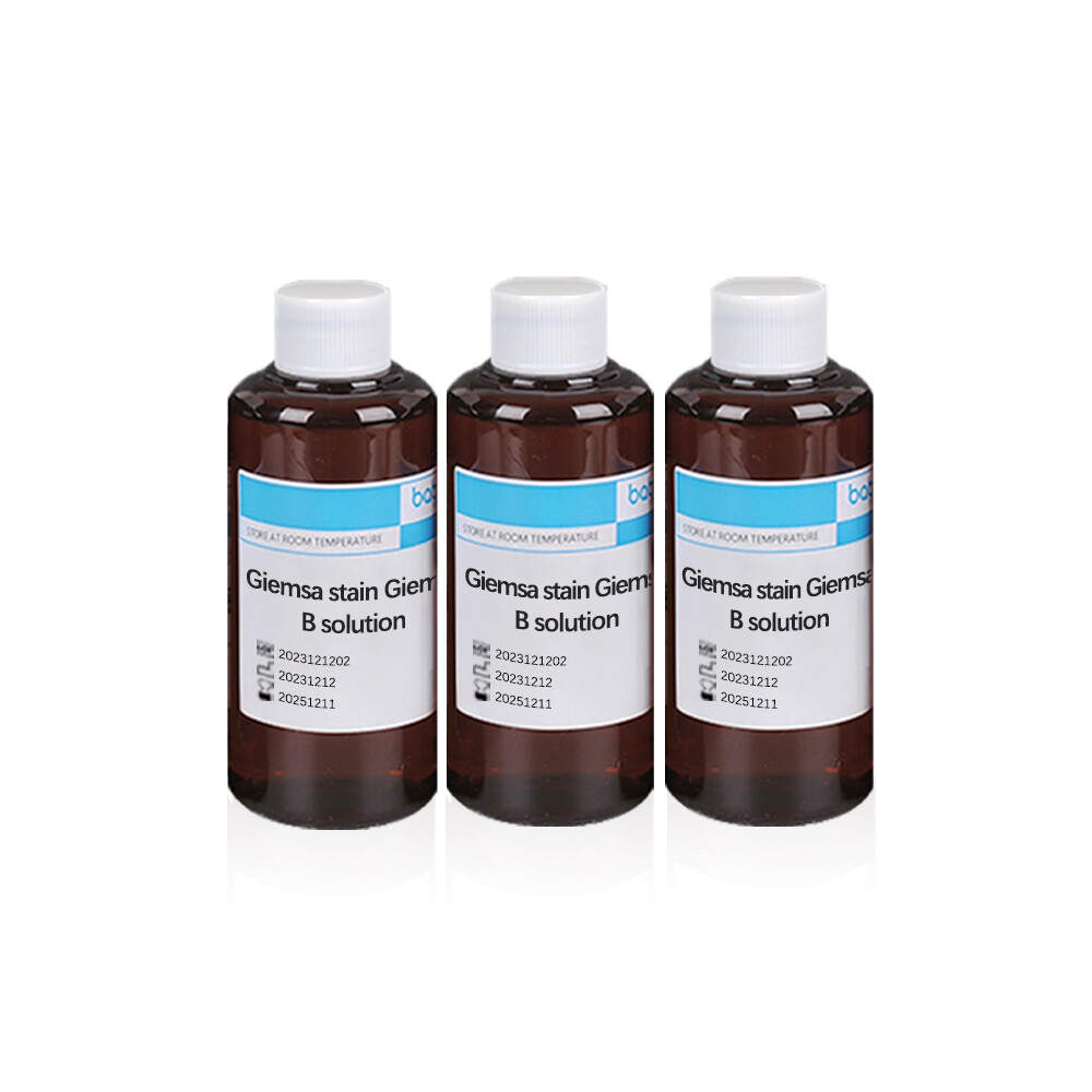
Baibo Biotechnology Co., Ltd., a leading Chinese manufacturer, presents its Giemsa stain Giemsa B solution. This high-quality Giemsa stain solution is ideal for lab tests, offering precise staining for various applications. The Giemsa stain kit includes a Giemsa stain buffer with a pH of 7.2, ensuring optimal results. Baibo Biotechnology’s expertise in producing reliable Giemsa stain solutions makes it a trusted choice for laboratories worldwide.
【intended use】
Giemsa stain Giemsa B solution mainly used for cell, chromosome and plasmodium smear staining
【principle】
Jimsa dye is a synthetic dye of Azul (blue)2 and eosin. Jimsa staining solution has strong staining power on cytoplasm and can better display the basophilic degree of cytoplasm, especially for azulocyan, eosinophilic and basophilic particles in blood and bone marrow cells, with clear coloring and pure color. The colored part of eosin is anion, the colorless part is cation, and the colored part is acidic. Methylene blue is usually alkaline chloride, the colored part is cation, colorless part is anion, just the opposite of eosin. The methylene blue is oxidized and contains azure blue, and the cell components are colored by selective adsorption of colored substances in it.
【Product specification】
A:1x20ml B:2x100m/ box;
A:1x100ml B:4x250m// Box:
A:1x250mlB:5x500m/ box
A:1x500ml B:1x5L/ box
【Operation procedure】
Hemocyte staining
① The blood cells are dried and fixed with methanol for 1-3 minutes:
(2) Place the fixed blood smear in Jemsa diluted staining solution (solution A: Solution B =1:9) for 10-30 minutes (less specimens can be stained by drops).
When the temperature is low, it can be dyed in the 37 degree temperature box:
③ Remove and rinse with water, dry and examine microscopically.
【Chromosome staining】
① The prepared specimens were treated with trypsin;
② Transfer to Jimsa diluted dyeing solution (liquid A: liquid B =1:9) for 10 minutes; ③ Wash it in water, dry it and examine it under microscope.
【Matters needing attention】
(1) The production of blood smears should be noted: the slide should be clean, the push and the slide should be maintained at a 30-45 ° Angle, and the blood should be waved in the air immediately after the push to dry it quickly;
The blood film is not dry, the cells are not firmly attached to the slide, and it is easy to fall off during the dyeing process, so the blood film must be fully dried:
(3) The concentration of the dye, the dyeing time and the temperature of the laboratory should be controlled, the lighter the dye, the lower the room temperature, the longer the dyeing time is required, so the dyeing time should be determined according to the specific situation; The dye should be added to cover the smear, not too little, so as not to evaporate and precipitate the dye;
④ When washing, the dye should be washed away with running water, and the dye should not be poured away first, so as not to deposit the dye on the blood sheet:
⑤ The dyeing solution can be reused, but it cannot be repeated countless times. If there is sediment, it should be filtered and used:
In order to obtain more white blood cells, the anticoagulant can be properly centrifuged, so that the cells with the same density are concentrated and stratified, and then the thin grayish white layer on the red blood cell layer (nucleated cells and platelets are concentrated) is smeated and stained. This method is very suitable for leukocyte classification and cell examination in patients with leukopenia.
【Result determination】
Blood cell staining: red blood cells are pink, red or orange-red: white blood cells are stained with different degrees of blue to dark blue nuclear chromatin structure is clear, cytoplasmic particles are clear, showing the unique color of various cells, such as neutrophilic granulocyte granules purple, eosinophilic granulocyte granules red, basophilic granulocyte granules purple
Chromosomal staining: On the chromosomes, different shades of horizontal pattern coloring can be displayed (deep band Jimsa coloring, light band coloring light or no coloring).
Online Message
Products Recommended


")


Please give us a message